
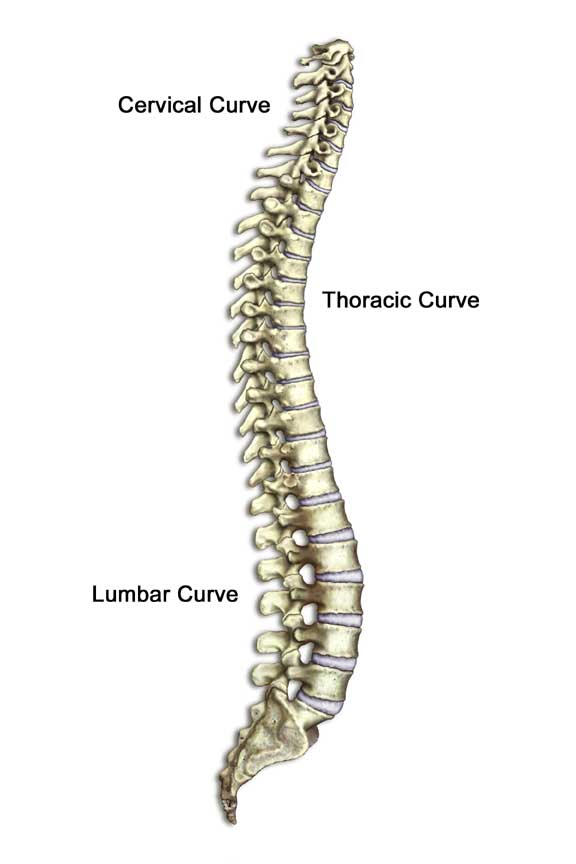
 Most people consulting their GPs for low back pain won’t be offered an X-ray, MRI or CT-scan, and other healthcare professionals would not advise these patients to go and see their GP to get a prescription for an X-ray.
Why is this?
85% of people with low back pain are diagnosed with “non-specific low-back pain” which means that there are no underlying diseases responsible for the problem. For that reason, there is no need to look for tissue damage with an x-ray as we know that no specific tissues are responsible for the problems.
For the other 15%, the history-taking and examination would reveal signs suggesting something more serious and possibly responsible for the problem. Investigations would then be offered.
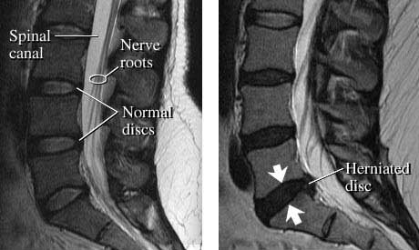 Twenty years ago most patients with back pain used to be offered X-rays. They had pain, an X-ray or MRI or CT-scan would be performed and signs of degeneration were seen leading to the conclusion that the patient’s pain was caused by the degeneration in the back. In the 90’s, studies started to look with X-rays, MRI and CT-scans at the backs of people with no back pain. Interestingly, they had pretty similar backs to patients with pain. The logical conclusion is that the changes seen on these images do not explain most patients’ pain (apart from the 15% mentioned above for which we know that investigations are necessary). Performing X-rays on everyone with back pain had three negative effects:
- unnecessarily irradiating patients who did not need an X-ray,
- a financial impact on the NHS,
- a nocebo effect. The nocebo effect is the opposite of the placebo effect in the sense that something that should be harmless causes symptoms. Patients started developing symptoms because they found out that there was some wear and tear in their spine and started believing that the pain was caused by this (which it is not!) This made it difficult to accept that the pain could improve, as the wear and tear would remain the same.
A systematic review of the literature (1) looked at all the studies on people with no back pain using either CT-scans or MRIs. Their conclusions are quite surprising.
In people with no back pain:
- nearly 4 out of 10 people aged of 20 years old have disc degeneration and 96% of 80-year-old individuals have it.
- 3 out of 10 twenty-year-olds have disc bulges and 8 out of 10 eighty-year-olds.
- 3 out of 10 twenty-year-olds had disc protrusions and more than 4 out of 10 eighty-year-olds.
- 2 out of 10 twenty-year-olds had signs of annular fissures and 3 out of 10 eighty-year-olds.
Sorry to repeat myself, but all of them were symptom-free! I’m not implying that disc degeneration / disc bulges / annular fissures are not painful, but most of us may have some discal changes that have already happened long before back pain. If we have discal changes but no pain, and later start having pain there may be another reason (or other reasons) than just what can be seen on an MRI or CT-scan!
Non-specific low-back pain is multifactorial (more information here) so we should not focus too much on MRI findings but more on how to improve the back’s function to get back to a normal life. There are several things we can do to help a bad back, including self-management (more information here) and seeking help from an osteopath of course! (more information here)
(1) W. Brinjikji, P.H. Luetmer, B. Comstock, B.W. Bresnahan, L.E. Chen, R.A. Deyo, S. Halabi, J.A. Turner, A.L. Avins, K. James, J.T. Wald, D.F. Kallmes, and J.G. Jarvik. Systematic Literature Review of Imaging Features of Spinal Degeneration in Asymptomatic Populations. AJNR Am J Neuroradiol 2014 Nov 27 [Epub ahead of print]
 Plantar fasciitis – should we stretch or strengthen?